At SmileArts Dental, we invest in advanced diagnostic tools to give patients clear answers and confident treatment plans. Cone-beam computed tomography (CBCT) has become an essential part of contemporary dental diagnostics because it delivers three-dimensional images that reveal structures a traditional X-ray simply cannot. This precision helps clinicians evaluate conditions with greater clarity, plan procedures more predictably, and communicate findings to patients in an understandable way.
Our CBCT technology is designed to balance image quality with patient safety. By producing targeted, high-resolution volume scans, we can visualize teeth, bone, nerve pathways, and sinus anatomy in fine detail while keeping exposures as focused as possible. The result is a faster diagnostic process, fewer surprises during treatment, and an overall improvement in clinical outcomes.
Traditional two-dimensional radiographs are helpful for many routine assessments, but they can obscure critical information when anatomy is complex or when structures overlap. CBCT provides volumetric images that remove those overlaps, revealing the true spatial relationships among teeth, jaws, and neighboring anatomy. This dimensional context is particularly useful when evaluating root morphology, tooth angulation, and the proximity of important anatomical landmarks.
The ability to rotate and slice through a captured volume means clinicians can examine areas of interest from multiple planes and at varying depths. Clinicians can quickly identify issues such as hidden canals, fractures, or bone loss that might be missed on a panoramic or periapical film. That depth of information supports more accurate diagnoses and reduces the need for exploratory procedures.
Beyond simply identifying problems, 3D imaging improves clinical confidence. When a diagnostic question is resolved by a CBCT scan, the care team can plan interventions with greater certainty. This clarity shortens the diagnostic pathway and helps avoid unnecessary delays, supporting smoother, more efficient visits for patients.
Accurate implant placement begins with a precise understanding of available bone volume and the position of vital structures such as nerves and sinuses. CBCT scans give a three-dimensional roadmap of the jaw, enabling predictable implant positioning and optimal prosthetic outcomes. Using these images, clinicians can determine implant length, diameter, and angulation before a single incision is made.
For oral surgery, CBCT is equally valuable. Impacted teeth, cysts, and lesions are mapped in relation to adjacent roots and anatomical boundaries, which helps the surgeon plan an approach that minimizes risk. The technology also supports template-guided surgery and digital workflows that translate virtual plans into physical guides for precise execution.
This preoperative insight reduces intraoperative uncertainty and can shorten surgical time, which contributes to a more comfortable experience and smoother recovery. In complex cases, CBCT data can be shared among specialists to coordinate care and confirm that every aspect of treatment is aligned with predictable goals.
Certain conditions demand a closer look than traditional imaging can provide. CBCT excels at detecting subtle changes in bone density, tooth root anatomy, and sinus health. It can reveal the extent of resorption, identify the course of root fractures, and uncover pathology that might be hidden from view on planar films. This more complete picture helps clinicians decide whether conservative treatment, endodontic retreatment, or surgical intervention is the most appropriate course.
For endodontics, CBCT is particularly useful in identifying root canal anatomy variations, locating missed canals, and assessing periapical lesions. In orthodontic and airway assessments, three-dimensional data clarifies relationships that influence treatment strategies, such as airway constriction or skeletal asymmetries. For sleep-related evaluations, CBCT can show craniofacial characteristics that may affect airway patency.
When pathology is suspected, CBCT imaging provides essential information that supports referrals and collaborative care. Clear documentation of findings helps clinicians communicate concerns to medical specialists and accelerates the path to a definitive diagnosis and treatment plan.
Patient safety is central to every diagnostic decision. Modern CBCT units use targeted fields of view and adjustable exposure settings so scans capture only the necessary anatomy. This approach limits radiation to the area of interest rather than imaging the entire skull, which aligns with established principles of radiation stewardship in dentistry.
Scans are quick to acquire, typically taking only a matter of seconds for the actual scan, which helps reduce the chance of motion artifact and increases patient comfort. The open, seated or standing design of many CBCT units also helps patients who find traditional enclosed imaging systems uncomfortable. The staff is trained to choose the smallest field and lowest exposure appropriate for each diagnostic question.
Clinicians interpret CBCT images in the context of the patient’s full clinical picture, including history and exam findings, so imaging is used judiciously. When a scan will meaningfully influence diagnosis or treatment planning, the benefits of three-dimensional visualization typically outweigh the small additional exposure compared with conventional films.
CBCT does not stand alone; it integrates with other digital tools to create a cohesive treatment process. Scans can be combined with intraoral scans and digital impressions to design restorations, surgical guides, and provisional prosthetics with precision. The resulting digital workflow shortens the time between diagnosis and delivery, and it improves the fit and function of final restorations.
Digital planning software allows clinicians to simulate procedures, evaluate outcomes, and make adjustments in a virtual environment before treatment begins. This capability supports predictable results and enables better patient education—visual aids derived from CBCT volumes help patients understand recommended treatments and expected outcomes.
In multi-disciplinary cases, digital CBCT files can be securely shared with specialists, enhancing collaboration and ensuring everyone involved has the same comprehensive information. This interoperability promotes coordinated care and helps streamline complex treatment sequences from start to finish.
In summary, cone-beam computed tomography is a powerful diagnostic tool that enhances accuracy, safety, and predictability across a wide range of dental treatments. If you’d like to learn more about how CBCT is used in our practice or whether a scan would be helpful for your dental care, please contact us for more information.


Cone-beam computed tomography, commonly called CBCT, is a three-dimensional imaging method used in dentistry to capture a volumetric view of the teeth, jaws, and surrounding structures. Unlike flat two-dimensional X-rays, CBCT produces a stack of images that can be reconstructed into axial, coronal and sagittal planes for detailed examination. This 3D perspective helps clinicians visualize spatial relationships and fine anatomy that are not always apparent on traditional films.
CBCT units capture the scan in a single rotation around the patient and generate a digital volume that can be manipulated with specialized software. The resulting dataset can be viewed as cross-sections, surface renders, or measurements to inform diagnosis and treatment planning. Because the images are digital, they are easy to archive, compare over time, and share with other providers when coordinated care is needed.
Traditional dental X-rays such as periapical or panoramic radiographs are two-dimensional projections that compress complex anatomy into a flat image. CBCT provides volumetric data so clinicians can inspect anatomy layer by layer, reducing ambiguity caused by overlapping structures. This dimensional information is especially helpful when evaluating root morphology, bone contours, and the proximity of nerves or sinuses.
Because CBCT produces a 3D dataset, measurements of bone height, width and angulation are more accurate than those taken from planar films. The improved visualization can reduce the need for exploratory procedures and support more predictable clinical decisions. However, CBCT is used selectively alongside conventional radiography depending on the diagnostic question.
A CBCT scan is recommended when three-dimensional detail will meaningfully affect diagnosis or treatment planning. Common scenarios include implant planning, evaluation of impacted or anomalous teeth, complex endodontic assessments, assessment of suspected fractures, and analysis of surgical anatomy near nerves or sinuses. It is also helpful for airway assessments and some orthodontic or temporomandibular joint evaluations when 3D relationships are clinically relevant.
At SmileArts Dental our clinicians consider the patient’s history, clinical exam and conventional radiographs before recommending a CBCT scan so imaging is used judiciously. The goal is to obtain the information necessary to guide care while avoiding unnecessary exposures. When a scan is expected to change the treatment approach or reduce intraoperative uncertainty, a CBCT is often the preferred option.
Modern CBCT systems are designed with radiation stewardship in mind and allow clinicians to select small, targeted fields of view and adjustable exposure settings. While CBCT exposes patients to more radiation than a single periapical film, the dose is typically lower than conventional medical CT and is justified when the diagnostic benefit outweighs the incremental exposure. Clinicians follow principles such as ALARA (as low as reasonably achievable) to minimize exposure while obtaining clinically useful images.
The scan itself is very quick, often only a few seconds of actual imaging time, which reduces the potential for motion artifacts and repeat scans. Protective practices include choosing the smallest field that answers the clinical question and using appropriate shielding when indicated. Your clinician will discuss the rationale for imaging and how it informs treatment so you can make an informed decision.
CBCT reveals three-dimensional relationships and internal anatomy that can be obscured on two-dimensional films, such as the exact course of a tooth root, accessory canals, root fractures, and localized areas of bone loss. It can delineate the position of impacted teeth relative to adjacent roots and important landmarks like the mandibular nerve or maxillary sinuses. This level of detail helps identify issues that might otherwise go undetected until treatment begins.
In addition to hard-tissue detail, CBCT can assist in assessing sinus anatomy, bone defects and the presence of lesions that require further investigation. The ability to slice through the volume at different angles aids in differentiating true pathology from artifacts or anatomic variations. These capabilities make CBCT a powerful adjunct when conventional imaging provides incomplete information.
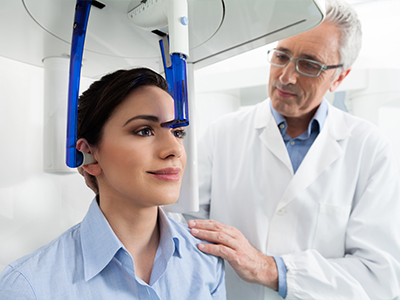
A CBCT scan is typically performed in an open, seated or standing unit depending on the device and patient needs. The patient is positioned so the head is stable and then the scanner rotates around the head while acquiring the images, a process that usually takes less than a minute. Patients are asked to remain still and may be given simple instructions such as biting on a small bite block or holding a steady posture to reduce motion.
There is no oral contrast or injection required for standard dental CBCT imaging and most patients experience only minor inconvenience during the brief procedure. After acquisition the images are reconstructed into a 3D volume and reviewed by the clinician using specialized software. The clinician will explain relevant findings and discuss how the images inform any recommended treatment steps.
CBCT provides accurate measurements of bone height, width and density, and shows the spatial relationship between planned implant sites and critical anatomy such as nerves and sinuses. This information allows clinicians to select appropriate implant dimensions and angulation before surgery, minimizing surprises and reducing the likelihood of intraoperative adjustments. Virtual planning based on CBCT volumes supports the fabrication of surgical guides that translate the digital plan into precise clinical execution.
For oral surgery, CBCT helps map impacted teeth, cysts and lesions relative to neighboring roots and anatomical boundaries so surgical approaches can be planned to limit trauma. The enhanced preoperative insight often shortens operative time and contributes to a smoother recovery. In complex cases, CBCT datasets can be shared with specialists to coordinate a multidisciplinary plan and confirm predictable outcomes.
Yes, CBCT is effective at revealing bony changes associated with infections, the presence and extent of periapical lesions, and certain root fractures that may not be clear on two-dimensional films. The three-dimensional views improve the clinician’s ability to localize pathology and assess how extensive a lesion is. This helps determine whether conservative therapy, endodontic retreatment, or surgical intervention is the most appropriate option.
CBCT can also reveal cortical bone destruction, resorption patterns and the relationship of pathology to adjacent structures such as the sinus floor or neurovascular bundles. While CBCT excels at visualizing hard tissues, soft-tissue details are limited compared with medical CT or MRI, so clinicians may recommend additional imaging or referral when soft-tissue characterization is required. Any suspicious findings are documented and, when needed, communicated to specialists for collaborative care.
CBCT datasets integrate with intraoral scans, digital impressions and CAD/CAM systems to create cohesive digital workflows for restorative, prosthetic and surgical treatments. Overlaying CBCT volumes with surface scans enables precise planning of implant positions relative to final prosthetic contours and facilitates the design of surgical guides and temporaries. This interoperability enhances the predictability of restorative outcomes and helps reduce adjustments at delivery.
Digital planning software also allows virtual simulation of procedures so clinicians can evaluate different approaches and communicate expected results to patients. The combined use of 3D imaging and digital design streamlines the path from diagnosis to restoration, often shortening treatment timelines and improving the fit and function of final prosthetics. Secure file sharing makes it straightforward to involve consultants or laboratories when multi-disciplinary input is required.
When a case benefits from multi-disciplinary input, CBCT volumes can be exported in standard formats and shared securely with specialists such as oral surgeons, endodontists, or radiologists for collaborative review. Sharing images helps ensure all providers work from the same detailed information and can coordinate a treatment plan that addresses each specialist’s concerns. This collaborative approach is particularly valuable in complex cases or when a second opinion will influence the care pathway.
The office of SmileArts Dental follows privacy and security practices when transferring digital imaging files and obtains patient consent for sharing when required. Clinicians will discuss with you why a referral or image-sharing is recommended and how it supports a safe, predictable outcome. If additional specialist input is suggested, the team will help coordinate the transfer of images and relevant clinical information to streamline communication.

Ready to Experience the SmileArts Dental Difference?
We’re here to help you take the next step toward a healthier, more confident smile. Whether you’re ready to schedule an appointment or want to learn more about our services, our friendly team is ready to assist you.
Our staff is happy to answer questions about treatment options, help you schedule your visit, and provide any information you need. Give us a call or fill out our quick online form today—we can’t wait to hear from you!