Digital radiography refers to modern x‑ray systems that capture dental images using electronic sensors and computer processing rather than traditional film. For patients, that translates into a faster, more streamlined experience: images appear on a monitor almost immediately, allowing your dentist to review findings with you during the same visit. The practical result is less waiting, clearer explanations, and the ability to move from diagnosis to treatment planning more efficiently.
At SmileArts Dental we use digital radiography as a routine part of diagnostic care because it supports clearer decision‑making while promoting patient comfort. The technology is flexible enough to capture a range of images — from small intraoral shots that show individual teeth to larger panoramic views that give a broad look at the jaw and surrounding structures. That versatility helps the clinical team spot issues early, track changes over time, and tailor care to your unique needs.
One of the immediate benefits patients notice is how the images are used during appointments. Rather than describing findings abstractly, clinicians can point to the same image you see on the screen, annotate it in real time, and illustrate treatment options visually. This shared view supports informed conversations and helps patients feel more confident about recommended care.
Compared with conventional film x‑rays, digital radiography significantly reduces radiation dose while maintaining or improving image quality. Because the sensors are more sensitive to x‑rays, they need less exposure to produce a diagnostic image. This reduction in radiation is a meaningful safety improvement, especially for patients who require routine imaging over the years.
Speed is another practical advantage: images are available instantly after capture, eliminating the delay of chemical development used in film systems. Faster processing means the dental team can evaluate concerns right away, which often shortens appointment times and helps avoid return visits solely for image review. From a clinical perspective, rapid access to images improves workflow without sacrificing diagnostic accuracy.
In addition to lower exposure and faster delivery, digital images can be enhanced with software tools that adjust contrast, brightness, and magnification. These tools help dentists detect subtle signs of tooth decay, bone loss, or other conditions that can be harder to see on traditional film, all while keeping patient safety a priority.
Digital dental imaging uses two main components: a sensor to capture the x‑ray and software to process and display the image. Intraoral sensors — thin, flexible panels placed inside the mouth — replace film for bitewing and periapical images. For broader views, extraoral sensors capture panoramic images or cone beam scans. Each type of sensor serves a specific clinical purpose and feeds high‑resolution data into the practice’s imaging system.
Once the sensor captures the image, specialized software converts the raw data into a clear, diagnostic picture. Clinicians can then apply enhancements, measure structures, and compare current and prior images within the same digital chart. This integration streamlines recordkeeping and supports longitudinal care by preserving high‑quality images in the patient’s file for future reference.
The combination of modern sensors and intuitive software also supports collaboration. When additional opinion or specialist input is needed, images can be shared electronically in secure formats, ensuring continuity of care without physical media. That digital exchange reduces the logistical friction of consultations and expedites coordinated treatment planning.
High-resolution digital images improve the dentist’s ability to detect small but important changes — from early cavities to subtle signs of infection or bone changes. Because images can be magnified and adjusted on screen, clinicians can examine fine detail without retaking exposures. Early detection often leads to simpler, less invasive treatment, which benefits both oral health and overall well‑being.
Digital radiography is especially helpful for treatment planning. Whether preparing for restorative care, endodontic treatment, or implant placement, clear images inform decisions about placement, angulation, and the extent of treatment needed. When combined with other in‑office technologies like intraoral cameras or 3D imaging, digital radiographs help create a coordinated, predictable clinical pathway from evaluation through completion.
Having consistent, high‑quality images in the patient record also supports follow‑up care. Progress can be tracked precisely over time, allowing the dental team to assess healing, monitor restorations, and catch new concerns early. That continuity improves outcomes and helps patients stay engaged in their own oral health.
Because digital images are stored electronically, they become part of a secure, searchable patient record that is easier to manage than paper film. Modern practices store images on encrypted systems that follow applicable privacy and security guidelines, minimizing the risk associated with physical media. Secure archives also simplify retrieval, so prior images are available for comparison during routine checkups or emergency visits.
Electronic storage makes sharing images straightforward when coordinating care with specialists or referring offices. Rather than mailing or hand‑delivering physical films, the practice can transmit files in secure formats that preserve image quality while protecting patient privacy. This rapid, reliable sharing reduces delays in diagnosis and supports a collaborative approach to more complex cases.
For patients, the visual nature of digital x‑rays enhances communication. Clinicians can walk through findings on‑screen, annotate areas of interest, and use the images to explain recommended steps. That visual dialogue often makes treatment options clearer and helps patients weigh choices with a better understanding of their oral health status.
Beyond technical advantages, digital radiography contributes to a more comfortable, patient‑focused experience. Smaller sensors, quicker appointments, and fewer repeat exposures combine to make imaging a less intrusive part of a dental visit. Staff can prioritize comfort while still collecting the detailed information clinicians need to provide excellent care.
The practice’s use of digital imaging is one way we strive to offer modern, evidence‑based dentistry in a friendly setting. Whether you’re visiting for a routine checkup or preparing for a specific procedure, the immediate access to clear images helps the dental team communicate more effectively and plan treatment with confidence.
For patients who appreciate transparency and clarity, digital radiography delivers both: better visuals to explain findings and a streamlined workflow that respects your time. If you have questions about how imaging fits into your care plan, the team is happy to explain the process and discuss what to expect at your next visit.
In summary, digital radiography is a safer, faster, and more flexible approach to dental imaging that enhances diagnosis, treatment planning, and communication. If you’d like to learn more about how this technology is used at SmileArts Dental or how it may affect your next appointment, please contact us for more information.


Digital radiography uses electronic sensors and computer processing to capture dental images rather than traditional film. Sensors convert X-ray exposure into digital data that appears on a monitor within seconds, allowing clinicians to evaluate findings immediately. This digital workflow eliminates chemical development and simplifies image storage and retrieval.
The practical difference for patients is faster appointments and clearer communication because the clinician can share and annotate images in real time. Image quality is often equal to or better than film, and digital files can be enhanced to highlight subtle findings. Overall, the technology streamlines diagnosis and supports more efficient treatment planning.
Digital radiography encompasses several image types, including intraoral bitewing and periapical images as well as extraoral panoramic views and cone beam scans for three‑dimensional assessment. Intraoral sensors record detailed views of individual teeth and supporting structures, while extraoral systems capture broader anatomic relationships. Each modality serves a different clinical purpose and is selected based on diagnostic needs.
Clinicians commonly use bitewings to evaluate interproximal decay, periapicals to assess root and periapical health, and panoramics to review jaw relationships and development. Cone beam imaging provides volumetric data for complex implant planning, impacted teeth, and some surgical evaluations. Choosing the right type of image helps ensure accurate diagnosis while avoiding unnecessary exposures.
Digital radiography typically requires less radiation than conventional film because modern sensors are more sensitive to X‑rays and produce diagnostic images with lower exposure. Practices follow the principle of keeping exposure as low as reasonably achievable and tailor imaging to each patient’s needs. Protective measures and careful technique further reduce unnecessary radiation.
Safety also depends on limiting repeat exposures by capturing high‑quality images the first time and using software enhancements instead of retakes. Clinicians assess risk factors such as age, medical history, and oral health to determine which images are necessary. If you have concerns about radiation, your dental team can explain why a particular image is recommended and what protections are in place.
Sensors capture raw X‑ray data and transmit it to specialized imaging software that converts the data into a viewable picture. Software tools adjust contrast, brightness, and magnification to help clinicians detect subtle signs of disease and measure anatomic structures. Integration with the patient’s digital chart keeps images organized for comparison and long‑term monitoring.
Advanced programs also allow annotations, measurements, and side‑by‑side comparisons with prior studies to support clinical decisions. When specialist input is needed, images can be exported in secure formats for timely consultation. This combination of hardware and software improves diagnostic accuracy and streamlines care coordination.
High‑resolution digital images make it easier to identify early cavities, bone changes, root fractures, and other conditions that benefit from prompt intervention. Clinicians can magnify areas of interest and enhance image parameters without additional X‑ray exposure, which aids detailed evaluation. Early detection often allows for simpler, less invasive treatments that preserve tooth structure and oral health.
For treatment planning, clear digital images inform decisions about restorative margins, endodontic anatomy, and implant placement. When combined with other in‑office technologies, such as intraoral cameras or 3D imaging, radiographs help create coordinated, predictable treatment pathways. Consistent imaging in the patient record also supports reliable follow‑up and outcome assessment.
Digital images become part of the patient’s electronic record and are typically stored on secure, encrypted systems that follow applicable privacy and security guidelines. Storing images digitally reduces the risk of lost or damaged film and enables quick retrieval for comparisons during future visits. Robust access controls and backup routines help preserve image integrity and availability.
When coordination with specialists or referring offices is needed, clinics transmit images using secure methods that maintain image quality and confidentiality. Electronic sharing eliminates the delay and fragility associated with physical media and speeds collaborative treatment planning. Patients may also be shown their images on screen to support clear discussions about care.
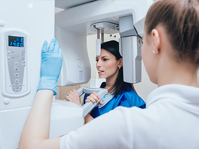
During an intraoral exam, a thin, flexible sensor is placed inside the mouth for brief exposures while the clinician or assistant positions the equipment. For extraoral panoramics or cone beam scans, you will be seated or asked to stand and remain still briefly while the machine rotates to capture images. The process is typically quick and causes minimal discomfort beyond sensor positioning.
After capture, images display almost immediately and the clinician can review them with you, point out areas of interest, and answer questions. If additional views are needed, the team will explain why and take steps to minimize exposure and maximize comfort. Clear communication throughout the visit helps patients understand findings and next steps.
The frequency of dental imaging is individualized based on age, oral health status, risk factors, and clinical signs rather than a fixed schedule. Patients with active disease, new symptoms, or high cavity risk may need more frequent monitoring, while low‑risk patients require fewer images. Your dentist performs a risk assessment to determine the appropriate type and timing of radiographs.
Routine recall exams often include selected images to compare with prior records and catch changes early, but the exact interval varies by patient. Clinicians balance the diagnostic benefit of imaging with the goal of minimizing exposure, and they will explain the rationale for any recommended images. If you have a specific concern about frequency, your dental team can review your history and preferences to set an appropriate plan.
Yes, digital radiographs integrate smoothly with other technologies such as intraoral cameras, digital impressions, and three‑dimensional imaging to create a comprehensive clinical picture. Combining modalities enables more precise treatment planning for restorations, endodontics, orthodontics, and implant dentistry. Software integration also allows clinicians to cross‑reference findings and simulate outcomes when appropriate.
This interoperability improves diagnostic confidence and helps the care team coordinate complex procedures with predictable steps. When specialists are involved, shared digital imaging expedites consultation and reduces logistical delays. The net effect is a more coordinated, evidence‑based approach to patient care.
At SmileArts Dental, digital radiography is a routine part of diagnostic care and is used to provide faster, clearer images that support informed conversations and efficient treatment planning. The office uses modern sensors and imaging software to capture a range of views, enhance diagnostic detail, and preserve high‑quality records for comparison over time. Immediate on‑screen review helps clinicians explain findings and involve patients in their care decisions.
The practice also leverages secure electronic storage and file sharing to coordinate with specialists when necessary and to maintain accurate, accessible patient records. By combining digital imaging with other in‑office technologies, the team strives to deliver predictable, patient‑focused outcomes while minimizing unnecessary exposure and appointment delays. If you have questions about imaging at your next visit, the staff in Yukon, OK is happy to walk you through the process.

Ready to Experience the SmileArts Dental Difference?
We’re here to help you take the next step toward a healthier, more confident smile. Whether you’re ready to schedule an appointment or want to learn more about our services, our friendly team is ready to assist you.
Our staff is happy to answer questions about treatment options, help you schedule your visit, and provide any information you need. Give us a call or fill out our quick online form today—we can’t wait to hear from you!