An intraoral camera is a compact, pen‑sized imaging tool designed to capture high‑resolution color photos and video inside the mouth. Unlike traditional mirrors, this device brings a magnified, illuminated view of teeth and soft tissues directly to a screen, giving both clinician and patient a clear and immediate visual of areas that are otherwise difficult to inspect. The result is a level of detail that supports more accurate assessments and a stronger shared understanding of oral health concerns.
These cameras are engineered to be comfortable and maneuverable, allowing clinicians to obtain close‑up views of molars, interproximal surfaces, and gum margins with minimal discomfort. The live feed can be paused to capture still images that document the condition of restorations, early caries, cracks, or soft tissue changes. Because the images are full color and high contrast, subtle differences in texture and shade become easier to evaluate than by sight alone.
For modern dental practices that prioritize clarity and patient engagement, intraoral cameras are an essential part of the diagnostic toolkit. They enhance the clinical exam rather than replace it, providing visual evidence that complements tactile exploration and other imaging methods, such as radiographs. In short, they translate what the clinician sees into something patients can see too.
One of the most valuable contributions of intraoral imaging is the ability to detect and document early signs of dental disease. High‑definition images reveal enamel demineralization, hairline fractures, margin gaps around crowns, and emerging periodontal issues that might be missed during a cursory visual exam. By capturing these findings at an early stage, clinicians can recommend appropriate preventive or restorative steps before problems progress.
Because the camera provides magnification and consistent lighting, it reduces the likelihood of overlooking minor but clinically relevant changes. The images can be compared over time to monitor lesion progression, restoration wear, or soft tissue healing. This longitudinal record supports evidence‑based decision making and helps clinicians prioritize interventions based on objective visual data.
When combined with other diagnostic tools—such as digital radiography and intraoral scanning—the camera’s images contribute to a more complete clinical picture. Each modality has strengths: radiographs show internal and bone structures, scanners create digital models for restorations, and the intraoral camera highlights surface detail and color. Together they inform a more accurate diagnosis and a more predictable treatment plan.
Visual information is a powerful communication aid. When patients can see a close‑up image of an area of concern, abstract explanations become concrete. The intraoral camera bridges the gap between professional observations and patient understanding, enabling informed conversations about oral health. Rather than relying solely on descriptions, clinicians can point to a specific photo and explain what it shows and why it matters.
This visual approach fosters trust and encourages active participation in care. Patients who can see evidence of plaque buildup, early cavities, or imperfect margins are more likely to grasp the rationale behind recommended preventive measures or treatments. It also helps set realistic expectations by showing the current condition rather than relying on hypothetical explanations.
At SmileArts Dental, we use intraoral images as part of our patient education process to make consultations clearer and more collaborative. Sharing visual findings empowers patients to make well‑informed choices about their care and to track improvements following treatment or hygiene changes.
Modern intraoral cameras interface easily with dental practice management systems and electronic health records, allowing captured images to be stored directly in a patient’s chart. This streamlined workflow preserves documentation for future visits and makes it simple to retrieve images for follow‑up discussions or referrals. Well‑organized visual records improve continuity of care when multiple providers or specialists are involved.
Images from intraoral cameras can also be shared with dental laboratories when fabricating restorations, giving technicians additional context about shade, contour, and existing anatomy. In restorative planning, photographs enhance communication about desired outcomes and provide reference points during the fabrication and try‑in stages. Visual documentation supports quality control and reduces back‑and‑forth by clarifying expectations.
For practices that utilize digital treatment planning—such as CAD/CAM workflows or implant planning software—intraoral photographs complement the data set by adding surface detail and color information that scans or radiographs alone may not capture. This integration leads to more precise, predictable results and a smoother experience for patients and clinicians alike.
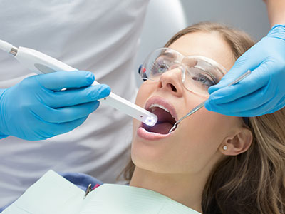
The use of an intraoral camera during a routine exam is quick, noninvasive, and tailored to each patient’s comfort. Typically, the clinician or dental hygienist will guide the small camera along the teeth and gums while displaying a live feed on a screen. The process usually takes only a few minutes and may involve capturing a handful of still images to document areas of interest.
Because the device is compact and designed for intraoral use, most patients find it unobtrusive. The clinician will review the images with the patient, point out observations, and explain any recommended next steps. If additional imaging or consultations are needed, the captured photos remain part of the patient’s record and can be referenced at future visits.
Patients benefit from the immediate visual feedback: seeing their own teeth magnified can motivate improved oral hygiene and make dental concepts easier to understand. The images also help patients verify the success of completed treatments by comparing before and after photos during follow‑up visits.
Wrap‑up: Intraoral cameras bring clarity to dental care by revealing small but important details, improving communication, and supporting thorough recordkeeping. If you’d like to learn more about how this technology is used during appointments or how it could benefit your oral health, please contact us for more information.


An intraoral camera is a compact, pen-sized imaging device that captures high-resolution color photos and video of the teeth and surrounding soft tissues. It provides magnification and dedicated lighting so clinicians can display detailed views on a monitor for both diagnosis and patient education. The camera is designed to reach molars, interproximal areas, and gum margins with minimal discomfort, making small defects and surface changes much easier to see.
Clinicians commonly capture still images from the live feed to document restorations, early decay, cracks, or soft tissue changes. Those images supplement tactile exams and radiographs by showing surface detail and color that other tools may not reveal. Overall, the intraoral camera translates professional findings into clear visuals patients can review and understand.
High-definition intraoral images increase the likelihood of detecting subtle problems that might be missed during a cursory visual exam, such as enamel demineralization, hairline fractures, and imperfect margins around crowns. The consistent lighting and magnification reduce variability between exams, enabling clinicians to spot minor but clinically important changes. Captured images can be compared over time to monitor progression or healing, supporting evidence-based decisions.
When used alongside radiographs and intraoral scanning, camera images add surface detail and color information to the diagnostic mix. This multimodal approach clarifies the clinical picture and helps prioritize interventions based on objective visual data. The result is more accurate treatment planning and fewer surprises during restorative procedures.
An intraoral camera exam is quick, noninvasive, and tailored to patient comfort; most sessions take only a few minutes to obtain a series of targeted images. The clinician or hygienist will guide the small camera along the teeth and gums while showing a live feed on a monitor and may capture a handful of still photos to document areas of interest. After imaging, the clinician will review the photos with you and explain any findings in plain language.
At SmileArts Dental we use intraoral imaging as part of routine exams to help patients understand conditions and treatment options more clearly. The images become part of your chart for follow-up reference and to demonstrate changes after treatment or improved home care. Patients often find the immediate visual feedback motivating for better oral hygiene and adherence to recommended care.
Yes. Intraoral cameras are designed specifically for intraoral use and are typically no larger than a writing pen, making them unobtrusive for most patients. The devices use visible light and safe imaging sensors rather than ionizing radiation, so they pose no radiographic risk. Clinicians and hygienists are trained in gentle handling techniques to keep the exam comfortable, even for patients with sensitive gag reflexes.
Many practices follow strict infection-control protocols and use barrier sleeves or clinically approved cleaning methods between patients. If you have concerns about comfort or sensitivity, discuss them before imaging so the clinician can adjust technique and positioning. The brief, targeted nature of the exam usually keeps discomfort to a minimum while yielding valuable diagnostic images.
Each diagnostic tool provides different, complementary information: radiographs reveal internal structures and bone levels, intraoral scanners create precise digital models for restorations, and intraoral cameras highlight surface texture and color. The camera’s color images help evaluate soft tissue health, margins, plaques, and small surface defects that scans or x-rays may not show clearly. Combining these modalities produces a more complete and reliable clinical assessment.
When clinicians integrate camera photos with radiographs and scans, they can cross-reference findings to improve accuracy and treatment predictability. For example, a suspicious area seen on an image can be correlated with radiographic evidence to determine whether restorative or periodontal intervention is needed. This coordinated approach reduces diagnostic uncertainty and supports better-informed treatment plans.
Intraoral cameras are highly useful for detecting early surface changes such as enamel demineralization, hairline fractures, and margin gaps that suggest restorative breakdown. The magnification and focused lighting reveal subtle textural and color shifts that are often precursors to more significant problems. Identifying these issues early enables clinicians to recommend preventive or minimally invasive treatments before larger restorations are required.
While camera images excel at surface detail, they do not replace radiographs for detecting subsurface decay or bone involvement. Clinicians will typically use cameras in conjunction with x-rays and clinical probing to confirm a diagnosis and determine the full extent of a lesion. This layered evaluation improves the chance of catching problems in an early, more treatable stage.
Modern intraoral cameras integrate with practice management systems to store captured images directly in a patient’s electronic chart, creating a permanent visual record. These images are tagged to specific dates and treatment areas so clinicians can track progression, document healing, and reference prior conditions at follow-up visits. Well-organized image records also support communication with specialists and laboratories when collaborative care is needed.
At SmileArts Dental we retain intraoral photos as part of each patient’s chart to ensure continuity of care and clear documentation of clinical findings. When appropriate, images can be shared securely with laboratories or referral partners to provide additional context for restorations or specialist evaluations. This documented visual history improves coordination and helps avoid unnecessary repetition of imaging.
Yes, intraoral cameras are commonly used with pediatric and adolescent patients because they are noninvasive and quick to use, making them suitable for routine exams and education. The visual feedback helps young patients understand oral conditions, which can improve cooperation and foster better daily hygiene habits. Clinicians adapt technique and positioning to a child’s size and comfort level to minimize anxiety and ensure clear images.
For very young or anxious children, clinicians may capture fewer images or focus on specific areas of concern while using a gentle, reassuring approach. Images can also be a helpful teaching tool for parents, showing plaque buildup, early decay, or areas that need closer monitoring. In this way, intraoral imaging supports preventive care and early intervention for younger patients.
Intraoral photographs provide accurate visual references for shade, contour, and the relationship of restorations to surrounding anatomy, which aids both clinicians and dental laboratories. These images can be sent with laboratory prescriptions to communicate expectations about margin fit, surface texture, and color nuances that influence the final restoration. During try-ins and adjustments, photos help verify that outcomes match the planned result.
When used with CAD/CAM scans and radiographs, camera images fill in surface and color detail that digital models and x-rays may not fully capture. This fuller dataset leads to more precise preparations, improved provisional restorations, and fewer adjustments at delivery. The visual documentation also helps clinicians explain each step of the restorative process to patients and set realistic expectations.
Intraoral images are treated as part of a patient’s protected health information and are managed according to privacy regulations and secure practice protocols. Practices typically store images within HIPAA-compliant practice management systems and limit access to the clinical team members involved in a patient’s care. Secure storage, controlled user access, and encrypted data transfer are common safeguards used to protect photographic records.
Patients who have questions about how their images are used or who they may be shared with should ask the practice staff for clarification before imaging. Clinicians will explain sharing procedures for referrals or laboratory communication and obtain any required consents. Clear policies and transparent communication help ensure that visual records are used appropriately and only for clinical or administrative purposes related to care.

Ready to Experience the SmileArts Dental Difference?
We’re here to help you take the next step toward a healthier, more confident smile. Whether you’re ready to schedule an appointment or want to learn more about our services, our friendly team is ready to assist you.
Our staff is happy to answer questions about treatment options, help you schedule your visit, and provide any information you need. Give us a call or fill out our quick online form today—we can’t wait to hear from you!