Wisdom teeth — the third molars at the very back of the mouth — are the last permanent teeth to form, usually emerging in the late teens or early twenties. For some patients they erupt without trouble and become useful chewing surfaces. For many others, however, wisdom teeth create issues because the jaw has limited space or the teeth develop at awkward angles. Recognizing what these teeth are and how they behave helps patients make informed decisions about monitoring or treatment.
Not all wisdom teeth cause symptoms right away. Some remain partially covered by gum tissue, while others are trapped beneath bone or lie tilted against neighboring teeth. These variations influence how a dentist or oral surgeon evaluates the situation and whether active intervention is recommended. Early identification through clinical exams and imaging is the first step toward preventing more serious problems down the road.
It is common for young adults and parents to wonder whether removal is necessary when X-rays show developing third molars. The answer depends on several factors, including the tooth’s orientation, the available space in the jaw, and the presence of infection or damage to adjacent teeth. A careful, evidence-based assessment avoids unnecessary procedures while ensuring problematic teeth are treated before they cause complications.
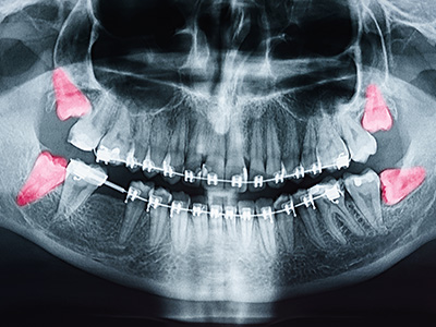
Evaluation begins with a thorough oral exam and a review of any symptoms a patient may have, such as pain, swelling, or difficulty opening the mouth. Visual inspection can reveal signs of gum inflammation or decay around a partially erupted tooth, but imaging is crucial for a complete assessment. Panoramic X-rays give a broad overview of tooth positions, while three-dimensional imaging (CBCT) offers precise details about root orientation and proximity to vital structures like nerves.
These imaging tools help clinicians classify impactions — for example, whether a tooth is soft-tissue impacted, partially erupted, or fully encased in bone. That classification influences treatment planning, anticipated difficulty of extraction, and postoperative recovery expectations. Imaging also identifies hidden problems such as cysts or resorption of adjacent tooth roots that may not be evident on clinical exam alone.
The evaluation process is collaborative: clinicians explain findings to the patient, discuss potential risks and benefits of treatment options, and outline the next steps. When a surgical extraction is likely, we coordinate care so patients understand what to expect during the procedure and the type of anesthesia or sedation that will be used based on comfort and safety considerations.
Impacted wisdom teeth can be the source of several oral health problems. When a tooth is trapped under gum tissue or bone, bacteria can accumulate around it, leading to recurrent infections and painful swellings. Partially erupted teeth create pockets where plaque and food debris collect, increasing the risk of gum disease and decay that can affect both the wisdom tooth and neighboring molars.
In some cases, pressure from an erupting wisdom tooth damages adjacent teeth or causes misalignment of the dental arch. Cysts can form around an unerupted tooth and slowly expand, weakening surrounding bone if left unchecked. Although tumors associated with third molars are uncommon, their potential presence is another reason clinicians take suspicious findings seriously and recommend appropriate imaging when concerns arise.
Even when a wisdom tooth is asymptomatic, progressive decay or periodontal problems might be developing out of sight. That’s why clinicians often monitor third molars over time and recommend extraction when the balance of evidence indicates a higher likelihood of future harm than benefit in retaining the tooth.
Removal is typically advised when a wisdom tooth is causing pain, infection, recurrent inflammation, or damage to adjacent teeth. In other situations, prophylactic extraction is considered when imaging shows that a tooth is unlikely to erupt properly and poses a predictable risk of future problems. The decision balances the risks of surgery with the likelihood of preventing more significant issues later on.
Age and timing play important roles. Extractions performed while roots are still developing — commonly in late adolescence and early adulthood — are often technically easier and associated with quicker recovery and fewer complications. That said, many adults come for evaluation later in life and still undergo safe, successful extractions; the approach is simply tailored to the individual’s anatomy and overall health.
When a treatment recommendation is made, clinicians discuss anesthesia options, potential risks, and realistic recovery expectations. If an extraction is deferred, regular monitoring with periodic exams and imaging ensures changes are detected early so an effective plan can be implemented without delay.
Wisdom tooth extraction may be performed under local anesthesia, sedation, or general anesthesia depending on complexity and patient preference. The goal is always to maintain patient comfort and safety. During the procedure, clinicians remove any overlying tissue or bone necessary to access the tooth and then extract it with careful technique that protects adjacent structures. Sutures may be placed to promote proper healing when indicated.
Following surgery, a structured aftercare plan helps minimize discomfort and supports healing. Patients are given instructions about rest, diet, oral hygiene modifications, and signs to watch for that would warrant prompt follow-up. Managing swelling, controlling bleeding in the immediate postoperative period, and maintaining cleanliness around the surgical site are key elements that reduce the likelihood of complications such as prolonged pain or infection.
While most recoveries follow an expected course, clinicians remain available for postoperative checks and to address questions or concerns. Clear communication about medication use, activity restrictions, and wound care improves outcomes and helps patients return to normal routines as safely and quickly as possible.
Once the immediate recovery period is complete, maintaining routine dental care is essential. Regular professional cleanings and exams allow the dental team to monitor healing, evaluate adjacent teeth, and manage any changes in gum health. Good daily oral hygiene — including careful brushing and flossing around the treatment area as advised — supports long-term stability and reduces infection risk.
For patients who retain some wisdom teeth, vigilant monitoring continues to be important. Even asymptomatic third molars can develop problems over time, so periodic radiographs and clinical assessments help detect changes early. For those who have had extractions, follow-up visits ensure bone and soft tissue heal properly and provide an opportunity to discuss broader preventive strategies.
Our approach is centered on evidence-based recommendations and clear communication so patients feel informed and confident in their care. If you have questions about the status of your wisdom teeth or would like a personalized evaluation, the team at SmileArts Dental is available to guide you through the options and next steps.
In summary, wisdom teeth can range from harmless to problematic, and careful evaluation with modern imaging and clinical expertise determines the best course of action for each patient. If you'd like more information about wisdom tooth evaluation or treatment options, please contact us for details and to schedule an appointment with our team.


Wisdom teeth, also called third molars, are the final permanent teeth to form at the back of the mouth. They typically begin developing in the late teens and often erupt between ages 17 and 25. While some wisdom teeth emerge fully and function without issue, others fail to erupt properly due to limited jaw space or awkward angulation.
These variations include fully erupted teeth, partially erupted teeth that remain covered by gum tissue, and teeth that are completely trapped beneath bone. Each scenario carries different risks and implications for oral health. Early identification through routine exams and imaging helps guide appropriate monitoring or treatment decisions.
Evaluation begins with a clinical exam to check for signs of swelling, infection, or decay and to review any symptoms a patient may have. Imaging is a central part of assessment; panoramic X-rays provide an overview of tooth positions while cone-beam computed tomography (CBCT) gives three-dimensional detail about root orientation and proximity to vital structures. This information lets clinicians classify impactions and anticipate surgical complexity.
Detailed imaging also reveals hidden problems such as cysts, root resorption of adjacent teeth, or atypical anatomy that could affect treatment. With clear diagnostic data, clinicians can explain risks and benefits and recommend the timing and type of care most appropriate for the individual. When necessary, advanced imaging helps tailor anesthesia and surgical techniques to improve safety and outcomes.
Impacted or partially erupted wisdom teeth can trap food and bacteria, leading to recurrent infections, gum inflammation, and painful swellings known as pericoronitis. They can also develop decay that is difficult to treat and lead to damage or increased risk of decay for neighboring molars. In some cases, pressure from erupting wisdom teeth contributes to crowding or changes in bite alignment.
Cysts may form around unerupted teeth and slowly expand, potentially weakening adjacent bone if not addressed. Less commonly, impacted third molars are associated with root resorption of neighboring teeth or the development of benign tumors, which is why suspicious findings are investigated promptly. Even asymptomatic teeth can harbor progressive problems that are best caught through monitoring and imaging.
Removal is recommended for wisdom teeth that cause pain, recurrent infection, persistent gum inflammation, or damage to adjacent teeth. Prophylactic extraction may also be considered when imaging shows that a tooth is unlikely to erupt properly and poses a predictable risk of future problems. The decision weighs the risks of surgery against the likelihood of preventing more significant issues later on.
Age and root development influence timing; extractions performed while roots are still forming—often in late adolescence or early adulthood—are generally easier and carry lower complication rates. That said, adults of any age can undergo safe extractions when clinically indicated, with treatment plans individualized to anatomy and overall health. When removal is deferred, regular monitoring ensures changes are detected and managed promptly.
Extractions range from simple removals under local anesthesia to more complex surgical procedures that may use sedation or general anesthesia for patient comfort and safety. The clinician removes any overlying gum tissue or bone necessary to access the tooth, then extracts the tooth with techniques designed to protect adjacent structures. Sutures are sometimes placed to help the tissues heal in an orderly fashion.
The team will review anesthesia options and explain intraoperative steps so patients know what to expect, including estimated procedure time and immediate recovery protocols. Safety measures include careful assessment of nerve proximity and surgical planning based on imaging findings. Clear communication before and during the appointment helps reduce anxiety and prepares patients for postoperative care.
Most patients experience the greatest discomfort and swelling in the first 48 to 72 hours after extraction, with noticeable improvement over the following week. Pain can usually be managed with prescribed or over‑the‑counter medications, ice packs, and rest, while soft foods and gentle oral hygiene help protect the healing sites. Stitches, when used, are often removed or resorb on their own within one to two weeks depending on the material.
Complete healing of the bone and soft tissue may take several months, although normal daily activities typically resume within a few days to a week for uncomplicated cases. The clinician schedules follow-up visits to confirm proper healing and to address any concerns that arise. Patients are advised to report increasing pain, fever, prolonged bleeding, or unusual swelling promptly so complications can be addressed early.
Following postoperative instructions carefully is the most effective way to reduce complications; this typically includes avoiding vigorous rinsing or spitting, refraining from smoking, and keeping the surgical area clean with gentle rinses after the initial 24 hours. Adhering to recommended activity limits and dietary guidelines helps protect the blood clot that forms over the socket, which is essential to healing. Taking medications as directed and keeping follow-up appointments also supports a smooth recovery.
Maintaining good oral hygiene while being gentle around the extraction site prevents bacterial buildup that can lead to infection. If signs such as increased pain after several days, persistent bad taste or odor, fever, or excessive swelling occur, patients should contact the dental team promptly for evaluation. Prompt attention to warning signs reduces the likelihood of more serious problems and speeds recovery.
Not all wisdom teeth require removal; when a third molar erupts fully, is functional, and poses no threat to neighboring teeth or periodontal health, conservative retention with regular monitoring may be appropriate. The decision to retain a wisdom tooth depends on its position, the patient’s oral hygiene, and the presence or absence of pathology on clinical exam and imaging. Periodic radiographs and clinical checks help ensure no new problems develop over time.
For retained third molars, the dental team assesses for early signs of decay, gum disease, or changes in surrounding structures and recommends intervention if risks increase. Patients who retain wisdom teeth should be diligent with routine cleanings and home care, and should report any new pain, swelling, or difficulty opening the mouth. Regular monitoring allows timely treatment should the status of a retained tooth change.
When third molar roots lie near important nerves—such as the inferior alveolar nerve in the lower jaw—detailed three‑dimensional imaging like CBCT is often used to assess the exact relationship and plan a safer surgical approach. Knowledge of nerve proximity can influence whether a tooth is removed in the office or referred for specialist care, and it guides choices about anesthesia and surgical technique. Surgeons use specific methods to minimize the risk of nerve injury while achieving the treatment goal.
In complex cases the team discusses the potential benefits and risks thoroughly so patients understand the likelihood of temporary or, rarely, permanent changes in sensation and the measures taken to reduce those risks. When appropriate, staged treatments or alternative approaches are considered to protect nerve function and overall oral health. Careful planning and advanced imaging are key to balancing efficacy and safety in these situations.
The team at SmileArts Dental emphasizes an evidence‑based, individualized approach that combines thorough clinical exams with modern imaging to guide decision making and treatment planning. Patients receive clear explanations of findings, anesthesia options, anticipated recovery, and follow‑up care so they can make informed choices with confidence. Sedation and comfort measures are offered when appropriate to ensure a positive experience during surgical procedures.
After treatment, follow‑up visits monitor healing, address any questions, and reinforce preventive strategies to maintain long‑term oral health. For patients in Yukon, OK and the surrounding communities, care is coordinated to provide timely evaluations and attentive postoperative support. If you have concerns about your third molars, scheduling an evaluation allows the team to assess your situation and recommend the best path forward.

Ready to Experience the SmileArts Dental Difference?
We’re here to help you take the next step toward a healthier, more confident smile. Whether you’re ready to schedule an appointment or want to learn more about our services, our friendly team is ready to assist you.
Our staff is happy to answer questions about treatment options, help you schedule your visit, and provide any information you need. Give us a call or fill out our quick online form today—we can’t wait to hear from you!